I have been debating this topic (creation vs. evolution) since Discover first opened up there Internet Site. An atheistic evolutionist, Gene90, and I would go rounds in the late 90’s on many of these topics. One of the topics that often came up as an evidence for evolution was JunkDNA, or, pseudogenes. They would argue, much like the vestigial organ argument, that there were upwards of 90% of the DNA that was vestigial, or useless. I would argue that the supposed vestigial organs were in fact useful, and the argument for JunkDNA was an argument from silence… much like the 180 vestigial organ list of the late 1800’s was. An argument — that upon elucidation — shed light on these many “vestigial” natures showing that in fact they had uses to our bodies. (My response to the vestigial organ argument follows this story.) Here again, the Design argument holds its place among the predictive powers that true science affords. This being said, philosophical naturalists reject this predictive power — NOT because it is scientifically unsound, but because an a priori understanding of these positions undermines their true application of science to their understanding of the natural world.
I was again recently in a debate on this topic thanks to some goofball on YouTube (beating a dead horse… I do this from time-to-time to get-to-know a subject well). I posted some of my responses to that elongated debate here: “The Vitamin-C Pseudogene Argument Crumbles Slowly.” (Also see especially: Pseudogenes and the Origin of Humanity: A Response to the Venema Critique of the RTB Human Origins Model) Which brings me to this post. Over at Debunking Atheists, there is an ICR update reproduced in part below. Take note that we, including myself in this grouping as an armchair apologist, have been saying this since the 90’s.
False Gene Discovery Confirms Creation Prediction, by Brian Thomas, M.S.
…Early analyses of all that “junk” revealed that it was not random, and later studies showed patterns in the non-gene-coding DNA, although at the time there were no known functions for them.1
A new study published in Nature disclosed the discovery of a totally new mechanism for gene regulation that uses pseudogenes.2 These look very similar to actual genes, but contain enough differences that they could not be used to properly code for proteins. For example, they often have a genetic “stop sign” buried in the middle of the sequence instead of at the end, where the coding gene has it.
The word pseudogene means “false gene.” They were named this because they were considered to be broken, useless copies of real genes that harbored coding errors from a long evolutionary history of genetic mistakes. But what if pseudogenes had those coding differences not because they were broken-down versions of the real genes, but because they were purposefully designed with specific similarities to help regulate their corresponding genes?
The researchers found exactly that in two gene-pseudogene systems. They documented direct evidence that the pseudogene PTENP1 “can regulate cellular levels of [its corresponding gene] PTEN and exert a growth-suppressive role.”2
Not only is the pseudogene PTENP1 not useless, it has two previously unknown functions. PTENP1 acts as a decoy to attract smaller regulatory molecules. When these small molecules attach to PTENP1 instead of PTEN, the PTEN gene becomes more accessible and is translated into protein more often.
The gene and its pseudogene also appear to exist in a balancing act that affects each other’s expression levels. This relationship was further established by observing that cells without the PTENP1 pseudogene are cancerous. Other diseases, like Cowden’s disease and Bannayan-Zonana syndrome, are associated with poorly regulated PTEN gene expression.3
The researchers suspected that this regulation scheme was not limited to just one pseudogene-gene system and that it might well be found throughout the living world. Sure enough, they found the same regulatory balances in the gene KRAS and its pseudogene KRAS1P.
So, a cell can regulate the output of genes and their protein products in “intricately dynamic” networks2 because of formerly designated “junk” DNA like transposons4 and pseudogenes. Since no networks–especially not those that provide essential regulatory functions–are seen arising from any natural process, these must have arisen from an intelligent and benevolent source outside of nature.
Based on the idea that both genes and non-gene DNA were produced by a Creator, rather than the laws of physics, creation scientist John Woodmorappe predicted in 2003 that pseudogenes would be discovered to play an important regulatory role in cells.5 Science is now showing that to be accurate.
References
1. The ENCODE Project Consortium. 2007. Identification and analysis of functional elements in 1% of the human genome by the ENCODE pilot project. Nature. 447 (7146): 799-816.
2. Poliseno, L. et al. 2010. A coding-independent function of gene and pseudogene mRNAs regulates tumour biology. Nature. 465 (7301): 1033-1038.
3. Keim, B. New Form of Gene Regulation Hints at Hidden Dimension of DNA. Wired Science. Posted on wired.com June 24, 2010, accessed June 29, 2010.
4. Thomas, B. Evolution’s Best Argument Has Become Its Worst Nightmare. Acts & Facts. 39 (3): 16-17.
5. Woodmorappe, J. 2003. Pseudogene function: regulation of gene expression. TJ. 17 (1): 47-52.
APPENDIX
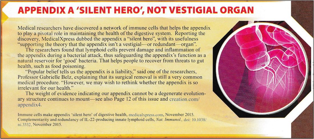
(Above graphic via Creation Magazine, Vol. 38 {No.2 – 2016} — click to enlarge)
- “There is no longer any justification for regarding the vermiform appendix as a vestigial structure.” William Straus, Quarterly Review of Biology (1947), p. 149.
- “For at least 2,000 years, doctors have puzzled over the function of… the thymus gland…. Modern physicians came to regard it, like the appendix, as a useless vestigial organ which had lost its original purpose, if indeed it ever had one. In the last few years, however,… men have proved that, far from being useless, the thymus is really the master gland that regulates the intricate immunity system which protects us against infectious diseases…. Recent experiments have led researchers to believe that the appendix, tonsils, and adenoids may also figure in the antibody responses.” – The Useless Gland that Guards Our Health, in Reader’s Digest, November 1966, pp. 229, 235.
- “The appendix is not generally credited with significant function; however, current evidence tends to involve it in the immunologic mechanism.” – 2Henry L. Bockus, M.D., Gastroenterology, 2:1134-1148 (chapter The Appendix, by Gordon McHardy), [W.B. Saunders Company, Philadelphia, Pennslyvania, 1976.]
- “The mucosa and submucosa of the appendix are dominated by lymphoid nodules, and its primary function is as an organ of the lymphatic system.” — Frederic H. Martini, Ph.D., Fundamentals of Anatomy and Physiology, p. 916, [Prentice Hall, Englewood Cliffs, New Jersey, 1995]
What Does It Do?
The appendix, in conjunction with other parts of the body which also contain cells called B-lymphocytes, manufactures several types of antibodies:
- IgA immunoglobulins, involved in surface or mucosal immunity. These are vital in maintaining the protective barrier between the bowel and the bloodstream.
- IgM and IgG immunoglobulins, which fight invaders via the bloodstream.
The appendix is in fact part of the G.A.L.T. (Gut Associated Lymphoid Tissue) system. The lymphoid follicles develop in the appendix at around two weeks after birth, which is the time when the large bowel begins to be colonized with the necessary bacteria. It is likely that its major function peaks in this neonatal period.
Dr. Kawanishi,[1] showed that human lymphoid cells in the appendix are immunologically functional as T helper cells and antibody-producing B cells, making IgA molecules in response to immunological challenges. He noted that:
- “The human appendix, long considered only an accessory rudimentary organ, could posses a similar antigen uptake role prior to replacement by fibrosed tissue after repeated subclinical infections, or at least in early childhood when it is most prominent.”[2]
The appendix is also rich in argentaffin cells, which can be identified with the use of silver salt staining. The function of these cells has long been obscure, but the evidence suggests that they may be involved with endocrine gland function.[3] Many sources (encyclopedias, textbooks, etc.) still erroneously state that the appendix is useless. Interestingly, the Grolier Multimedia Encyclopedia states in one place: that “In humans the cecum and appendix have no important function,” and in another place that “the appendix is now thought to be one of the sites where immune responses are initiated.”
Dr. Howard R. Bierman… studied several hundred patients with leukemia, Hodgkin’s disease, cancer of the colon and cancer of the ovaries. He found that 84% [of his sample] had [their] appendix removed…. In a control group without cancer, only 25% had it removed.[4]
Bierman himself had concluded that the appendix may be an immunological organ whose premature removal during its functional period permits leukemia and other related forms of cancer to begin their development.[5] Bierman and his associates realized that the lymphoid tissue located on the walls of the appendix may secrete antibodies which protect the body against various viral agents.
While high school and college textbooks today will mention the appendix as vestigial, specialists in their field have for many years stated the necessity of the appendix as useful.
- “There is no longer any justification for regarding the vermiform appendix as a vestigial structure.”[6]
- “For at least 2,000 years, doctors have puzzled over the function of… the thymus gland…. Modern physicians came to regard it, like the appendix, as a useless vestigial organ which had lost its original purpose, if indeed it ever had one. In the last few years, however,… men have proved that, far from being useless, the thymus is really the master gland that regulates the intricate immunity system which protects us against infectious diseases…. Recent experiments have led researchers to believe that the appendix, tonsils, and adenoids may also figure in the antibody responses.”[7]
- “The appendix is not generally credited with significant function; however, current evidence tends to involve it in the immunologic mechanism.”[8]
- “The mucosa and submucosa of the appendix are dominated by lymphoid nodules, and its primary function is as an organ of the lymphatic system.”[9]
The appendix is in fact part of the G.A.L.T. (Gut Associated Lymphoid Tissue) system. The lymphoid follicles develop in the appendix at around two weeks after birth, which is the time when the large bowel begins to be colonized with the necessary bacteria. It is likely that its major function peaks in this neonatal period.
As Dr. Peter Faletra (Ph.D.), who is Senior Science Advisor Office of Science Department of Energy, says in response to a question on an online question-and-answer service for K-12 teachers run by the Argonne National Laboratories:
“As a histologist I see no reason to consider the v. appendix as having no function since it contains numerous lymphoid follicles that produce functional lymphocytes and a rich blood supply to communicate them. The general idea of vestigial organs is to me a measure of ignorance, arrogance and lack of imagination. Ignorance in that we label it as such because we do not know its function; arrogance in that we declare it of no value since we can see none; and lacking in imagination in so far as when we cannot see its function cannot imagine one. I call your attention to that other ‘vestigial organ’ the thymus without which, in early life, we would produce a severely compromised cell-mediated immune system as the ‘nude’ mouse and numerous thymectomized mammalian studies have shown. Although some general reference books still list the v. appendix as ‘vestigial’ most immunologists (I included) would strongly disagree!”[10] (emphises added)
Footnotes
[1] H. Kawanishi, “Immunocompetence of Normal Appendiceal Lymphoid cells: in vitro studies,” Immunology, 60(1) (1987), pp. 19-28.
[2] Ibid., p. 19.
[3] Marti-Ibanez (editor), “Tuber of Life,” M. D. Magazine (1970) #14, p. 240; William J. Banks, Applied Veterinary Histology (Williams and Wilkins, Baltimore: 1981), p. 390.
[4] Richard G. Culp, Remember thy Creator (Baker Book House, Grand Rapids,; MI: 1975).
[5] Howard R. Bierman, “Human Appendix and Neoplasia,” Cancer 21 (1) (1968), pp. 109-118.
[6] William Straus, Quarterly Review of Biology (1947), p. 149.
[7] “The Useless Gland that Guards Our Health,” in Reader’s Digest, November (1966), pp. 229, 235.
[8] Henry L. Bockus, Gastroenterology, 2:1134-1148 [chapter The Appendix, by Gordon McHardy], (W.B. Saunders Company, Philadelphia, Pennslyvania: 1976).
[9] Frederic H. Martini, Ph.D., Fundamentals of Anatomy and Physiology, (Prentice Hall, Englewood Cliffs, New Jersey: 1995), p. 916
[10] From the site Newton, which is an electronic community for Science, Math, and Computer Science K-12 Educators. Argonne National Laboratory, Division of Educational Programs, Harold Myron, Ph.D., Division Director. Quote: http://www.newton.dep.anl.gov/askasci/mole00/mole00225.htm Home page: http://www.newton.dep.anl.gov/